Any medical manipulations during the bearing of a child inspire doubt in women. Hence, when receiving the next direction, the question arises: is it possible to do an ECG during pregnancy? Women can be understood, since recently they have become responsible not only for their lives, but also for the lives of their children. Therefore, even such a harmless procedure as an ECG during pregnancy requires a serious and thoughtful approach.
What is an ECG?
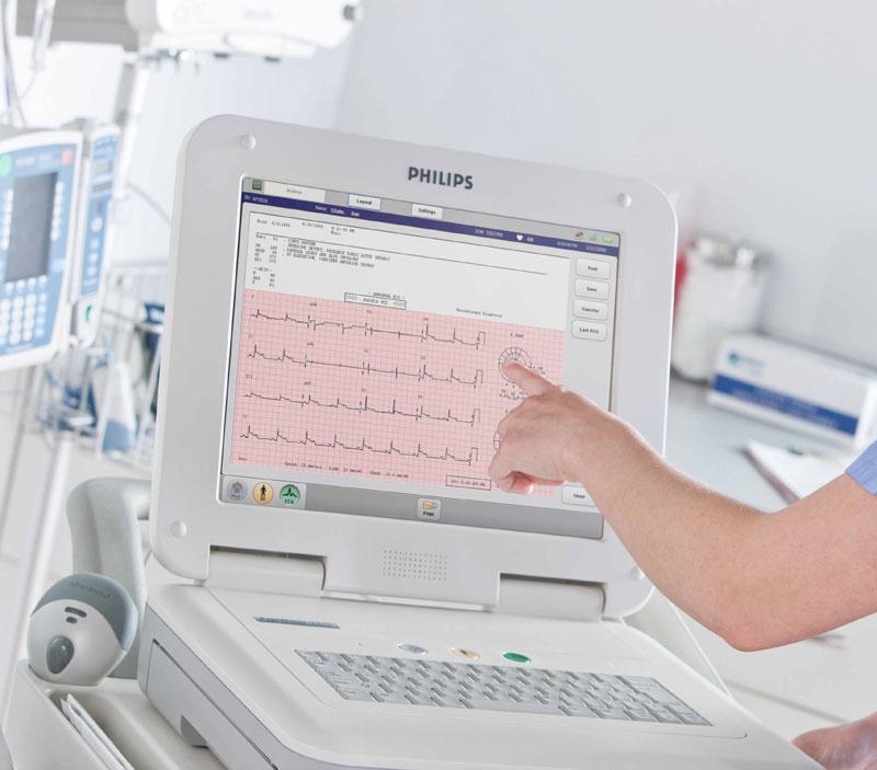
Electrocardiography is a method of studying the electric fields that are formed as a result of the work of the heart. The abbreviation ECG stands for "electrocardiogram", which, in turn, is a printout obtained from a study of the heart muscle.
The ECG procedure is a fairly inexpensive, but very informative diagnostic method in cardiology. It is carried out using special devices, which, receiving pulses through the electrodes, record them on thermal paper. Modern electrocardiographs allow you to immediately save the ECG of patients in digital form without listing.
What diseases can be detected by ECG?
Electrocardiography allows you to identify many diseases and pathologies of the heart. It is prescribed during routine medical examinations, as well as in early pregnancy. An ECG can detect:
- Violations of intracardiac patency.
- Diseases associated with impaired heart rate (arrhythmia, extrasystole).
- Myocardial damage.
- Disruptions in the exchange of electrolytes (potassium, calcium, etc.).
- Some extracardiac diseases, such as obstruction of the pulmonary artery.
- Acute cardiac abnormalities.
As a rule, electrocardiography is included in the mandatory list of examinations for medical examination. In addition, an ECG during pregnancy can be prescribed unscheduled if a woman has indications for this procedure.
Indications
During pregnancy, a woman’s heart muscle begins to work with a vengeance. This is due to the fact that the saturation with oxygen and beneficial substances of the fetus occurs through the blood. In addition, during this period, the level of hormones rises, which also affect the functioning of the heart.
An ECG during pregnancy is usually prescribed in the first trimester. It is included in the list of recommended studies, especially if:
- a woman has constant jumps in blood pressure;
- there are complaints of sharp or dull pain in the region of the heart;
- a pregnant woman suffers from headaches, dizziness, fainting;
- there are pathologies of gestation (polyhydramnios, gestosis, etc.)
During the entire period of pregnancy, healthy women undergo electrocardiography once. There are no contraindications for recording an ECG during pregnancy, so it is prescribed to absolutely all women who are registered with a health care institution.
ECG preparation
As with any procedure, it is advisable to prepare for electrocardiography. This is necessary to quickly and easily go through the study without the need for re-recording.
Recommendations for preparing for the ECG:
- For the procedure, it is better to choose clothes that can be easily unfastened on the chest.
- On the appointed day, you can not apply creams and other cosmetics to the skin, as they can disrupt electrical conductivity.
- In the decollete zone there should not be chains, pendants and other jewelry that would interfere with fixing the electrodes.
- Just before the study, you need to tell the doctor about all the medications that are currently being used, especially heart ones.
Also, if a woman is going to undergo an ECG during pregnancy, she needs to avoid physical activity immediately before the procedure. Therefore, going up to the office by the stairs, there is no need to rush. But if nevertheless there is shortness of breath before entering electrocardiography, the pulse is increased due to physical fatigue, you need to sit a bit and wait until the heart rate recovers and returns to normal.
How do ECGs during pregnancy?
Electrocardiography takes place in health facilities - clinics, hospitals, medical centers. Today there are portable devices with which a doctor can record an ECG even at home. However, so far they are used only for patients who are not able to independently get to a medical facility.
The standard ECG removal procedure is as follows:
- The patient exposes the area of the chest, forearms, legs and fits on a special couch.
- The doctor applies a gel to these areas, which reduces electrical resistance.
- The electrodes are attached to special points on the body where the greatest electrical conductivity is. During the examination, they will transmit pulses to the device, which will translate them into a graphic image.
- During recording, the patient should breathe calmly evenly. Your doctor may ask you to take a deep breath and hold your breath for a while. The patient must follow the instructions silently, since talking during the ECG is prohibited.
- In order for the ECG to be as informative as possible, the patient's body should be at rest. Movement and even involuntary trembling can “smear” the real ECG results.
- After the recording is completed, the electrodes are detached, the gel residues are wiped from the skin. The result of the ECG is transmitted to the doctor, who gave a referral for examination.

This procedure is quite simple. As a rule, it takes no more than 5-7 minutes. But the abundance of electrodes usually scares women and makes one doubt whether an ECG is possible during pregnancy.
Contraindications
Caring for the health and development of the child, before giving consent to medical examinations, women are primarily interested in the available contraindications. In the case of electrocardiography there are none. Absolutely all doctors, including gynecologists, say that you can do an ECG during pregnancy, regardless of the patient's condition. The only side effect that can occur after the procedure is a rash in the places where the electrodes are attached. As a rule, this is an individual intolerance to the gel, which is used during the examination. However, such rashes are not dangerous. They pass through 1-3 days on their own.
ECG Results Analysis
Decipher the evidence obtained after electrocardiography, only a doctor can. It takes an average of 10-15 minutes for experienced specialists, after which the results of the ECG are transmitted to the gynecologist, who issued a referral for examination.
In the conclusion, which is given by the result of electrocardiography, it is indicated:
- heart rate pattern;
- heart rate (heart rate);
- the electrical axis of the heart muscle;
- the presence or absence of conductivity disturbances.
If the ECG was prescribed according to available indications, then for the diagnosis, the doctor analyzes the combination of symptoms and signs of the disease. In the most serious cases, the patient can be hospitalized for a complete and thorough examination.
Features of the cardiogram in pregnant women
During pregnancy, the nature of the cardiovascular system changes dramatically. She begins to work for two, and this, in turn, cannot but affect the ECG. The difference is especially noticeable when examining women in the third trimester of pregnancy.
The cardiogram of a pregnant woman is characterized by:
- Displacement of the electrical axis of the heart muscle to the left.
- Heart rate increase.
- Reducing the PR interval.
- An increase in the depth of the Q wave in the third lead and in all pectorals on the right.
- The T wave consists of two leads, it can also become both positive and negative.
These changes are explained by an increase in cardiac output, which is typical for pregnant women. This physiological feature arises from the need to ensure normal blood flow in the placenta and the fetus. Also, the features of the cardiogram in pregnant women are affected by weight gain and a shift in the position of the heart in the chest. Therefore, in order to avoid making an erroneous diagnosis when decoding the ECG, the doctor must take into account the position of the patient.