For any woman in position, there is nothing nicer than hearing the fetal heartbeat. And what could be better than a sound symbolizing the emergence in the female body of a new life ?! But what’s interesting is, at what period can you feel this wonderful music of a small heart? Let's try to figure it out. Typically, the heart rate (HR) can determine the gender of the unborn child. But let's not get ahead of ourselves, we will consider everything in order.
The first treasured sounds
By 2 or 3 weeks of fetal development in the womb, a heart begins to form in it. But so far it is a simple tube. For this reason, the expectant mother may not even know that she already carries a new life under her heart, which is at the initial stage of development.
After another two weeks, the tube acquires an S-shape, because of this, this stage in the development of the children's heart is referred to as sigmoid. After another 4-5 weeks, a septum forms inside the organ, resulting in three chambers. Someone may immediately ask the question: "And when does the heart begin to beat in the fetus?". That's right, starting from this moment, the little heart begins to make its first contractions.
During the first trimester, the general condition of the fetus is assessed by heartbeat. During listening, three main characteristics are identified:
- Heart rate.
- Rhythm.
- The nature of the heartbeat.
You can only hear these sounds using the transabdominal method using special sensors. But if there is no special indication for this, then it is better to abandon this manipulation. And by the end of 5 months of pregnancy, the baby's heartbeat can be heard through a conventional medical stethoscope.
Need for listening
The child’s heart is auditioned for a reason, and there are good reasons for this. And above all, this concerns the establishment of the fact of pregnancy. As soon as a woman has a delay in the menstrual cycle, the first thing she thinks is that it is necessary to do the appropriate test. And with a positive result, many ladies go to the hospital in order to perform their first ultrasound.
When the heart begins to beat in the fetus, we have already found out, now it’s worth understanding why it is necessary to listen to it. But it is not always possible to detect a heartbeat, which is not yet a pathology. It will certainly make itself felt, but a little later. It is worth worrying in those cases when, during the re-examination, nothing is still heard. This may indicate that the fetal egg is deformed, which is not good. Often in this case, a frozen pregnancy is diagnosed, in which an abortion should be done in connection with medical indications.
In addition, the heartbeat allows you to assess what condition the fetus is in the womb. In this case, the body is able to feel everything that is happening in its environment. When the expectant mother is experiencing stress, she has some kind of ailment, or she exposes herself to excessive physical exertion, the oxygen saturation of the fetus decreases. As a result, this is reflected in the form of abnormalities in the fetal heart rate. But, as a rule, such changes are temporary, and the increased work of a small heart is usually due to a violation of the blood supply to the fetus, which is called fetoplacental insufficiency. Often, this condition becomes chronic, and therefore does not lead to compensatory changes.
In addition, the heartbeat allows you to assess the condition of the child before childbirth. During this process, he and his heart are exposed to enormous loads: squeezing, a small amount of oxygen. At the physiological level, the child’s cardiovascular system has already been prepared, having acquired resistance to stress during the entire period of pregnancy.
However, sometimes the umbilical cord can be pinched, or in case of placental abruption, there is a serious danger. There may also be other no less threatening conditions. For this reason, obstetricians vigilantly monitor the baby's heartbeat after each fight.
Ways to Listen to the Heart
Before we touch on the norms of the fetal heart rate, we will consider how exactly you can listen to it. Usually, various devices are used for this. Moreover, depending on the gestational age, one or another diagnostic technique is used:
- Ultrasound
- Echocardiography (ECG).
- Auscultation.
- Cardiotocography (CTG).
Ultrasound scan
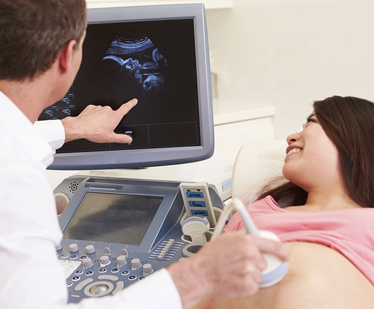
Ultrasound examination is the very first method that is used to assess the condition of the fetus. Moreover, starting from the first month of pregnancy. At an earlier date, a transvaginal (vaginal) examination is performed, and in a later period, the transabdominal method (through the stomach) is used.
This method allows you to identify various pathologies, and in the early stages of pregnancy. For the entire time a child is born, a woman needs to undergo a study at least three times.
Already on the first visit, you can find the first heartbeat of the fetus. During the second visit, you can view his camera, which will reveal the presence of defects or other deviations. If the doctor creeps in some suspicions, then he prescribes an additional study, during which all 4 cameras will be “visible”. As a result, doctors identify up to 75% of the likely pathological conditions of the children's heart.
In the period of 2 and 3 trimesters with the help of ultrasound, the volume of the organ and its position are determined. Under normal conditions, the heart is located in the region of a third of the volume of the chest.
Echocardiography, or ECG
As a rule, this study is prescribed for pregnant women in the event that several violations were detected during the ultrasound:
- delayed fetal development in the womb;
- deviations in the work of the heart;
- pathological condition of the fetus;
- abnormalities in the structure of the heart.
An ECG allows you to evaluate not only the fetal heartbeat, but also the structure of the main organ of the circulatory system, and in detail: does it perform all the functions, and are there any blood flow disorders in all its chambers. For this, one- and two-dimensional images, dopplerometry are used. To obtain a reliable result, it is better to conduct such a study from 18 to 28 weeks according to the obstetric period of pregnancy.
In addition, an ECG can be prescribed to a woman after she reaches 38 years of age, in the presence of any endocrine disease (diabetes mellitus), cardiovascular damage (CHD or congenital heart disease). In addition, if the expectant mother during the pregnancy suffered a disease of an infectious nature, or gave birth to children with CHD, the gynecologist can also prescribe her an echocardiography.
Auscultation
This technique is applicable with the onset of 5 months of pregnancy. Its essence lies in listening to heart rhythms with a stethoscope through the surface of the abdomen. The procedure is carried out not only at every visit by a woman to a gynecologist. Immediately during childbirth, every 20 minutes, the obstetrician is listening to the fetal heartbeat in order to assess the condition of the child.
In addition, the doctor can determine his position in the womb:
- Head presentation - in this case, the heartbeat can be heard below the navel.
- The transverse position of the fetus is indicated by listening to heart rhythms at the level of the navel.
- Pelvic presentation is detected when listening to heart beats above the navel.
In addition, during the auscultation, you can determine the rhythm and nature of the heartbeat. Which in turn implies the possibility of identifying not only hypoxia, but also pathologies in development.
At the same time, such a procedure in some cases may become ineffective:
- When the placenta is located on the front wall of the uterus.
- With a large amount of amniotic fluid or, conversely, oligohydramnios.
- Multiple pregnancy.
- The woman is obese.
But despite this, auscultation is considered a fairly reliable and easy to use method for measuring fetal heart rate.
Cardiotocography, or CTG
This technique is based on the registration and collection of analyzes of the work of the heart muscle under various conditions, with or without movement, during uterine contractions, against the background of various stimuli. In the presence of oxygen deficiency, the method is able to detect such a condition without problems.
The danger of hypoxia, which is oxygen deficiency, is to reduce the adaptive capabilities of a very young organism, which often results in a slowdown in its development and growth. As a result, there is a high risk of various pathologies during and after childbirth.
By means of CTG, two parameters of the fetal heart rate are determined:
- heart rate variability;
- basal rhythm.
The term "basal rhythm" refers to heart rate during the movement of the child and in its absence. Normal heart rates are 109-159 beats per minute at rest and 190 when moving.
As for the rhythm variability, this is the difference in heart rate between the state of rest and during movement. With normal development, the parameter should be 5 to 25 contractions, no less and no more. Any deviations from the norm are considered pathology. At the same time, relying only on these values, such conclusions should not be made, since additional studies are needed.
Varieties of CTG
When prescribing cardiotocography, it can be conducted in one of two ways:
- External (indirect) research.
- Internal (direct) research.
With indirect diagnosis, the fetal heartbeat and uterine contractions are examined using special sensors placed on the stomach. This method does not have any contraindications and can be used not only during pregnancy, but also during direct birth.
As for direct diagnosis, it is used in the rarest cases and only during the birth of a child. The study is carried out using several devices: an ECG electrode, which is attached to the baby’s head, and a sensor inserted into the uterus.
The result is evaluated by a special point system. 9-12 is considered the norm. 6-8 points indicates a mild hypoxia, as a result of which the woman will have to undergo a second examination the next day. 5 - this is already pronounced oxygen starvation, which poses a serious threat to the baby (or baby). In this case, you have to give birth only by caesarean section.
Heart rate indicators by week
What is characteristic, the fetal heartbeat is uneven over the weeks of pregnancy, and is gradually accelerating each time. Initially, the work of the heart is similar to the maternal rhythm. But subsequently, heart rate begins to increase, which is due to the accelerated formation of the body crumbs. The greatest frequency of muscle contractions occurs at 9-10 weeks of pregnancy, but then it falls.
By the arrival of 14-15 weeks, the main organs and their systems have already been formed, in the future they only grow. By the last term, heart rate can vary from 130 to 160 beats per minute. For clarity, the figure below shows the normal fetal heart rate by week.
With the onset of 12 weeks of bearing a child by heart rate, you can determine its gender:
- Less than 140 beats per minute - a boy will be born.
- More than 140 beats per minute - a girl will appear.
Thus, it can be noted that in girls the heart works much more intensively than in boys. At the same time, the heart rhythm is also different: again, in the male half it is measured, while in the female half it is more chaotic.
Possible deviations
We have already familiarized ourselves with the normal indicators of the work of the children's heart from the table above. But in some cases there may be serious deviations. So, changes in heart rate may indicate the following conditions:
- Tachycardia. This condition can be caused by insufficient circulation of the uterus and placenta, a small amount of hemoglobin in the mother's circulatory system, fetal anemia, placental insufficiency, placental abruption. Also, a rapid heartbeat of the fetus can be observed due to the pathological condition of the heart, the high temperature of the expectant mother, the inflammatory process of the membranes, the use of drugs like atropine or ginipral, an increased level of intracranial pressure of several other factors.
- Bradycardia The development of this condition contributes to the long position of the expectant mother on her back. This causes compression of the inferior vena cava. but besides this, there may be other reasons: treatment with propranololom, heart defects.
All of the above should not be underestimated due to the seriousness of the situation. For such reasons, a woman needs proper treatment, and in some cases it is impossible to do without the use of a caesarean section.
Finally
Finally, it remains to wish each expectant mother to monitor her condition, especially during pregnancy. It is necessary to strictly observe all the recommendations of the doctor who leads the whole process. In particular, this applies to ultrasound and other necessary and additional procedures.
It is no coincidence that during each planned ultrasound, the doctor listens to the baby’s heartbeat. His heart rate, rhythm and the nature of heart contractions can tell a lot about things to a specialist. Sometimes you can even determine the gender of the fetal heartbeat. It is clearly not worth ignoring such examinations if a woman wants to hug her full and, most importantly, healthy child!