The day is approaching faster when the expectant mother becomes real and sees her long-awaited baby. The decisive third trimester comes when the social status of the baby officially changes. Now he becomes a child from the fetus.
Third trimester. What happens to the baby?
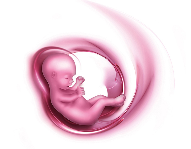
The third trimester lasts from 28 to the fortieth week and will be marked by the active growth and development of the baby. During this period of time, the child begins to accumulate subcutaneous fat and becomes more and more like a newborn. Already at 32 weeks, he will reach a weight of about 1.8 kg and will be about 28 cm in length. Before giving birth he will gain more body weight up to 3-3.5 kg, he will form wakefulness and sleep cycles, and he will begin to suck his thumb pens, preparing to suck on the mother’s breast. In the third trimester comes the finish line. Now your baby is becoming more active, smiles and frowns, trains respiratory functions and prepares to enter the big world.
Ultrasound When do they do it?
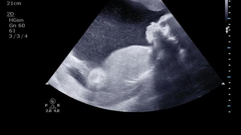
This period is the most informative. Therefore, an ultrasound of the fetus is carried out in the third trimester. And at this time not only the usual ultrasound examination is prescribed , but also the planned mandatory third screening. This next examination is very important to assess the current state of the fetus and its position before the onset of labor. In the third trimester, in what week will the doctor appoint an ultrasound? As a rule, local gynecologists send the expectant mother to a planned regular ultrasound examination at about 30-33 weeks. But in some cases, it can be carried out according to indications and in the periods from 28 to the thirtieth week, and at 34-36 weeks.
What does an ultrasound show? What pathologies can be detected?
Ultrasound in the third trimester is a mandatory procedure for every pregnant woman. It is completely painless, but it makes it possible to identify possible fetal pathologies at an early stage or to obtain final confidence in the child’s excellent health. In addition, this procedure allows you to determine the weight of the baby in the womb, as well as its gender. Moreover, ultrasound of the fetus in the third trimester allows us to find out the exact dimensions of the fetal head and its torso. It also turns out to evaluate the condition of the placenta and determine the exact position of the fetus in the uterus.
Ultrasound data of the third trimester is unique information that accurately shows all measurements, norms and possible deviations from them, which only a qualified specialist can decrypt. Based on the results of such an examination, the doctor makes a decision regarding the general health of the woman and her fetus. if necessary, prescribes additional studies or gives directions for hospitalization. If there are any deviations from the norm, ultrasounds in the third trimester will help to detect them and specify them with the help of an additional examination. In this period of pregnancy, a Doppler study of the vessels of the fetus and umbilical arteries is indicated. Since their work is very important for the cardiovascular system of the future crumbs.
In addition, ultrasound in the third trimester allows you to determine whether the fetus receives enough nutrients and oxygen to exclude the development of hypoxia and other cardiological pathologies. The information obtained gives an expanded idea of the course of pregnancy and the intrauterine development of the unborn child. These indicators are important not only for the doctor, but also for ensuring the calmness of the expectant mother. But if the period allotted for the third trimester of pregnancy is fourteen weeks, then when is the best time for a planned study? In the third trimester, which ultrasound week shows more accurate and reliable results?
Screening
The best time to carry out a planned screening ultrasound is considered to be 30-32 weeks. It was at this time that there was already enough information about all the necessary parameters that, according to the norms, the fetus should reach, as well as the condition of the placenta and uterus. In addition, since the child at this time becomes more active, one should pay attention to the location of the fetus, where its arms, legs, head are located, is the fetus lying correctly and are there any pathologies in its organs. Therefore, those who are interested in the question of when they do an ultrasound in the third trimester can answer that the most effective period is 30-32 weeks. Although you can make it in 29 weeks, but then everything will be more blurry and difficult to distinguish. When the testimony of the study is fuzzy, it is difficult to track the appearance of genetic abnormalities and the development of the baby’s organs, and it’s not always possible to clearly determine its gender. As a rule, women try to do it at the 30th week to do an ultrasound in the third trimester. The terms are already such that they allow you to consider everything thoroughly, but it is still far from giving birth.
What points do you pay special attention to during ultrasound?
At this time, attention is paid to such moments as:
- The position in which the fetus is in relation to the uterus of the mother. If it is located upside down, then there is no cause for concern, the child is lying normally, occupying the correct position. But it often happens that the unborn child is spread across and the doctor gives him a period of 2-3 weeks to take a normal position. If a coup did not happen during this period, mom will be prepared for cesarean section so as not to harm either the baby or his parent.
- The sufficiency of amniotic fluid, because it is when they do an ultrasound in the third trimester, you can find such a deviation from the norm as low water or high water. Both the first and the second are very dangerous for expectant mothers, because it signals the presence of any infections in the body.
- The entwining of a child with an umbilical cord is a fairly common deviation, and at this time you can even determine a double entwining. If the fact of entwining the umbilical cord is confirmed by ultrasound, then only a caesarean section is recommended by specialists - in the process of natural birth, the child can simply be strangled with his own umbilical cord while passing the birth canal.
- The degree of maturation of the placenta - if it has matured before the due date, corresponding to the stage of development of pregnancy, then the woman should be constantly monitored so that premature contractions and childbirth do not begin, moreover, with early maturation of the placenta, the child will experience a deficiency of nutrients and oxygen.
- Only an ultrasound in the third trimester allows you to most accurately determine the weight of the unborn baby, which is of great importance with a narrow pelvis of a pregnant woman, when the doctor has doubts about whether she can give birth on her own.
- Fetometry. These are the parameters for measuring the volume of the fetus - the head, abdomen, thigh length, because it is precisely on these indicators that the gestational age is established. Having discovered deviations, the doctor is obliged to carry out an extended procedure of phytometry - he measures the circumference of the head in the frontooccipital part and considers its percentage with other measurements. Then he makes a repeated measurement of the tummy and compares it with the measurement of the femur. After the measurements, the doctor examines the brain, examining the state of the plexus of blood vessels, the size of the lobes of the brain and cerebellum, which is required to check for brain diseases and intrauterine infections that can negatively affect the motor and swallowing capabilities of the child. After that, the doctor examines the structure of the nose, lips, eyes and spine.
- The condition of the fetal organs - especially the lungs and heart. If its diaphragm is underdeveloped, it means that the lungs will also not correspond to the norm. To check the cardiac activity, the correct operation of the valves, blood vessels and septa, a special study is conducted - cardiotocography, which allows you to determine the heart rate and check all the cardiac activity of the system. This procedure can only be performed after 32 weeks, otherwise the diagnosis will provide false data.
- Status of the abdominal cavity - the coordination of the intestines, liver, kidneys and bladder is checked. Of the pathologies, deviations in the kidneys most often occur.
Is ultrasound harmful to the baby in the womb?
At 30-32 weeks, an ultrasound is performed by simply driving the sensor along the wall of the abdomen of a pregnant woman. This is a completely harmless procedure, since the ultrasonic waves used in the device do not harm either the future, the mother, or her fetus. This is especially important for those who are interested in how often to do ultrasound in the third trimester. Since today ultrasound is the most effective and safe way to conduct a high-quality diagnosis during pregnancy, the recommendations of doctors in such cases should not be neglected. Only this research method is able at an early stage to identify potential pathologies and reduce the risk of their occurrence even before the birth of the baby.
Almost every woman who often has had an ultrasound scan in her third trimester worries about whether she has harmed her unborn baby. But do not worry about it. Since it has been proved by medicine that at this frequency, on which the devices work, no harmful effects occur either on the pregnant woman herself or on her unborn child. This is an absolutely routine procedure, which is prescribed by a doctor at a later stage of pregnancy, if you need to follow the development of a particular organ of the fetus. To assess blood circulation, dopplerometry is used, which studies the vascular network, placental blood flow and the cardiac function of the child in more detail.
Norms of indicators and measurements of the fetus
If the doctor prescribed an ultrasound in the third trimester, what week is the best time to perform fetometric measurements and what is their norm? Possible deviations from the norm in the development of individual organs of the child may indicate physical underdevelopment of the fetus. Control measurements of various fetal parameters are made in the period from 32 to 34 weeks. They should in normal condition correspond to such indicators:
- biparietal head size - 78-82 mm plus or minus 7 mm;
- fronto-occipital part - 104-110 mm plus or minus 9 mm;
- head circumference - 304-317 mm plus or minus 21-22 mm;
- tummy coverage - 286-306 mm plus or minus 28-30 mm;
- the length of the femur is 61-65 mm plus or minus 5 mm, the bones of the lower leg are 56-60 mm plus or minus 4 mm, the humerus is 56-59 mm plus or minus 4 mm, the bones of the forearm are 49-52 mm plus or minus 4 mm.
According to the state of the placenta - its location, thickness, structure, degree of maturity, various important points are clarified: if the placenta is located close to the uterus, the risk of fixing the head in an incorrect state may develop. The thickness of the placenta can vary from 32.2 mm up to 43.8 mm, if there is a mismatch in the parameters, then the function of entering the body of the fetus of nutrients is disrupted. The structure of the placenta should be as uniform as possible. Otherwise, there is a high probability of the development of any inflammatory process.
Uterine amniotic fluid should have an exclusively vertical diameter and be in a free area with sizes from 20 to 70 mm.
Pathologies that can be detected at a given time
In the third trimester, pregnancy is already moving towards its successful conclusion and this period has the following possible pathologies that are detected by ultrasound:
- incorrect fetal position;
- deviation in the amount of amniotic fluid;
- entwining the baby with an umbilical cord;
- degree of placental maturity;
- mismatch of the parameters of the fetometric measurement;
- pathology of the heart, lungs and abdominal organs.
Doctor's advice to expectant mothers
Gynecologists recommend that pregnant women strictly follow the instructions of the leading doctor and pay attention to their physical condition and psychological state. After all, the third trimester brings a lot of inconvenience to the life of the expectant mother, caused by an increase in the size of the uterus, fear of an impending genus, lower back pain, displacement of internal organs, shortness of breath, frequent urination, periodic constipation, varicose troubles. In addition, there is a feeling of anxiety and fear. Doctors recommend that during this crucial period, carefully monitor that the state of health was normal. If signs of gestosis or placental abruption are found, or any other problems with the body, contact a medical institution for help. Only the well-being of the expectant mother can serve as a true indicator of the favorable course of pregnancy. Any deterioration should be considered as an occasion for a visit to the doctor.
Little conclusion
Now you know when to do an ultrasound examination of a pregnant woman in the third trimester. We examined all the norms of indicators. Possible pathologies that can be seen on an ultrasound scan were also called. We hope that this information will help you make a healthy baby. Having reached all the features of the course of the third trimester of pregnancy, you can more carefully listen to your body, which will certainly warn you about an imminent meeting with your baby.